What Are The 3 Types Of Dental Implants
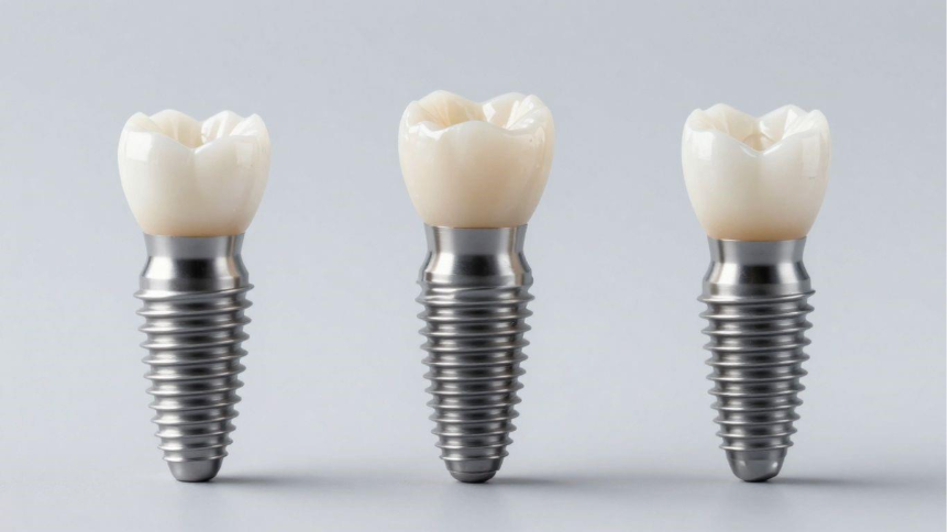
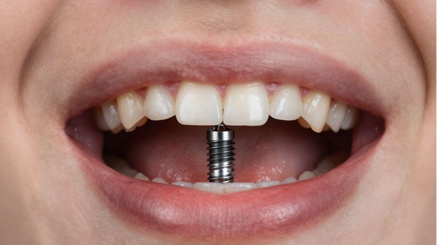
Missing teeth can really affect how you feel about your smile, and sometimes even how you eat or talk. Dental implants are a great way to fix that, giving you back strong, natural-looking teeth. But figuring out which kind to get can be a bit confusing. This post is here to break down the main 3 types of dental implants, so you can feel more confident about your choices for your oral health.